Hydronephrosis & Antenatal Counselling

Currently most pregnant women undergo one or two ultrasound scans during their pregnancy. When the fetus is scanned, kidneys and the urinary tracts are analyzed, In addition the volume of liquor (amniotic fluid) also helps to suspect kidney problems. Of late stretching of the kidney (Hydronephrosis) is detected very frequently.

Kidney Stretching (Hydronephrosis)

During scanning of 100-200 pregnant ladies, one fetus is found to have this abnormality. This usually happens because of a blockage in the urinary tract. Before proceeding further it is essential to describe the normal arrangement of the kidneys.

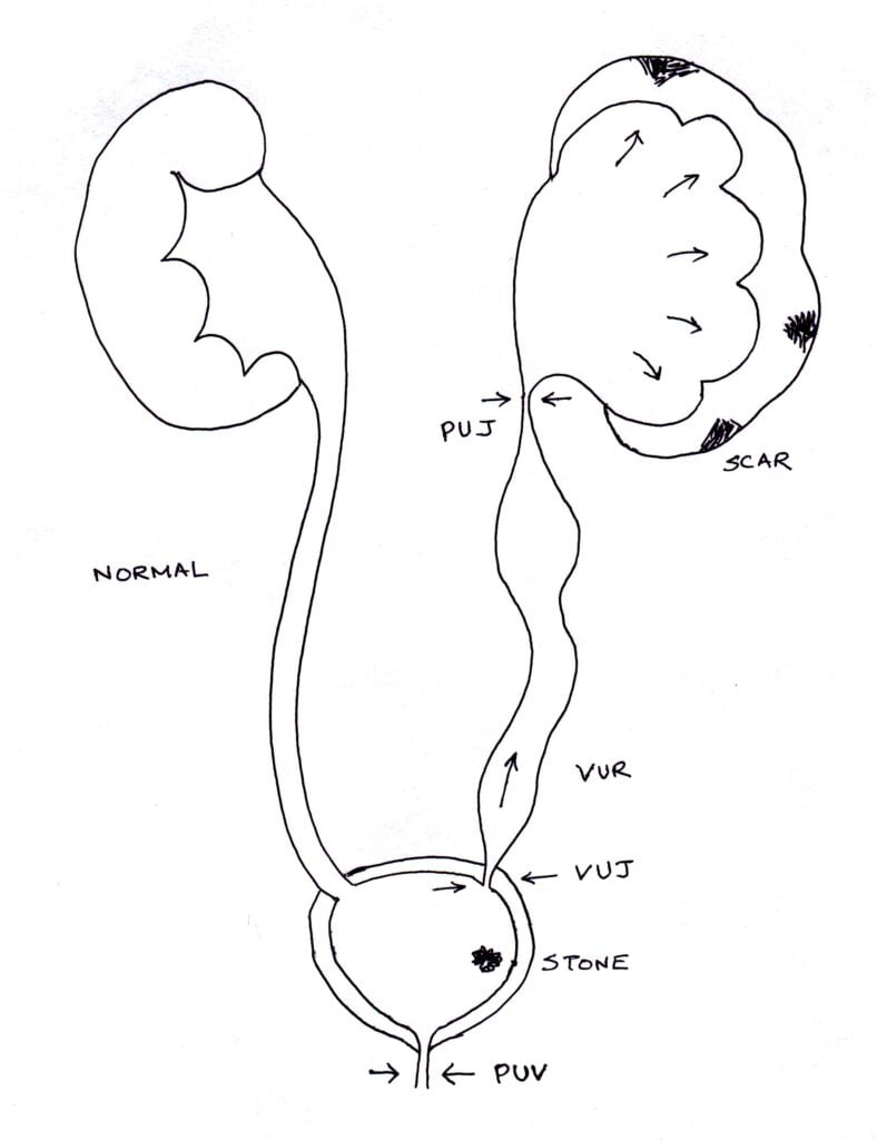
In human body there are two kidneys, one on either side of belly. From each kidney urine drains out via a tube called ‘ureter’ into a common urinary bladder. The bladder is located in the lower most part of belly. Once enough urine accumulates, it empties via a tube called ‘urethra’. Whenever there is a block in any of these tubes, kidney stretching results.

hydro

Single Kidney Stretching (Unilateral Hydronephrosis)

When only one kidney (right or left) stretches, blockage is suspected only on that side. When kidneys only stretch, the blockage is suspected at a higher location (PUJ obstruction). When the pipe draining the kidney also stretches, blockage is suspected at a lower location (VUJ obstruction).

Newborns with this problem need to be scanned on the third day after birth. In general very mild stretching is likely to settle spontaneously. Huge stretching requires surgery urgently to prevent loss of kidney function (renal failure). Children with moderate stretching need close monitoring with repeat scans and further tests. It is essential to consult a pediatric urologist to determine what tests are required and when a surgery could be avoided. Also it is essential to prevent urinary infection by taking a small dose of antibiotic once at night.

Both Kidneys Stretching (Bilateral Hydronephrosis)

When both kidneys stretch, they can be blocked separately. But usually they are blocked as urine exits the bladder in the ‘urethra’. In boys a valve like blockage can result in stretching of both kidneys (posterior urethral valve – PUV). Immediate endoscopic intervention after birth is essential to prevent urine infection and kidney failure.

Consultation before birth (Antenatal Counseling)

When there is a fetal kidney problem, there could be a lot of anxiety among parents. Consulting a paediatric urologist can relieve the anxieties and also provide answer to many questions. It will help to plan the type of delivery, place of delivery (where the treatment is available to the baby after birth), the type of tests required after birth. Also it helps to find out more about what is expected in the long run (prognosis) and whether the problem can occur again in future children (familial incidence).

Key Points:

  • It is possible to determine kidney problems in the baby before birth.
  • Most of the kidney problems correct spontaneously, although it is essential to confirm this by consulting a Paediatric Urologist
  • A small proportion of babies require corrective surgery after birth.
  • By consulting before delivery, parents can relieve their anxieties.

Hydronephrosis (PUJ)

This section discusses about blockage caused by PUJ obstruction. In this condition there is a blockage of urine at the junction of kidney and the pipe draining (ureter) – pelvi ureteric junction (PUJ). Because of this the kidney stretches. Often it affects one side alone although it can involve both kidneys.

What are the symptoms of hydronephrosis?

  • Detected before birth (Antenatal Scans): The condition should not cause the mother to have any symptoms. Hydronephrosis should not cause the baby to have any problems before birth, but he or she needs close monitoring after birth to keep an eye on the condition.
  • Colic or Pain abdomen: This can be a feature in older children. This is because of the urine in the kidney trying to push and overcome the blockage.
  • Urinary Infection (UTI) Whenever there is any blockage to urine flow, the system can easily catch infection. These children may have fever, frequent painful urination etc.

How is hydronephrosis diagnosed?

  • Ultrasound Scan: Hydronephrosis is diagnosed using an ultrasound scan. It helps to determine the severity of blockage; reflected by the amount of dilatation.
  • Nuclear Scan: For this test a small dose of medicine is injected in to the vein; pictures are taken with a gamma camera (like x-ray) every few minutes; this shows how fast the kidneys take up the medicine and how fast it leaves the kidney. When there is a blockage, the medicine stays in kidney for a long time. When the kidney function is poor; very little medicine is taken up by the kidney.
  • MCU: This is mainly to exclude problems like reflux or a valve.

How can hydronephrosis be treated?

  • If the hydronephrosis is diagnosed during pregnancy, early treatment will consist of monitoring with ultrasound, to check that the baby is growing normally and the kidneys are not getting too large. The baby will usually be born by a routine delivery.
  • After the baby is born, the hydronephrosis will be monitored using ultrasound scans and Nuclear scans.
  • When the hydronephrosis is getting worse or the kidney function is reducing surgery called pyeloplasty is recommended.

What does a pyeloplasty involve?

Under general anaesthesia, a small cut is made in the kidney region. The blocked portion is removed and reconnected. A small tube called stent may be kept to protect the sutures. It may be kept fully inside (needs removal under a second anaesthesia) or partly out (bedside removal after 7 days). The hospital stay is between 4-8 days. Children recover very well and it is not very painful as medicines will be given to take care of it. They can have normal feeds within 24 hours of operation.

What is the outlook for children with hydronephrosis?

  • If the cause of the hydronephrosis is an obstruction and this is removed shortly after birth, kidneys will be able to work properly.
  • The earlier the hydronephrosis is discovered, the better the outcome.