Pregnancy Planning, Anomaly Scan Prevention of fetal anomalies
Pregnancy Planning
In addition to social and financial factors, there are several other reasons for planning pregnancy. One such is planning pregnancy from the point of view of preventing anomalies. Organ formation in the fetus is completed by 5-8 weeks following conception. However a lady often detects pregnancy only after this (pregnancy test is often done when periods are skipped for more than 6-8 weeks), thereby missing the golden chance to prevent anomalies.

Planning pregnancy helps one to take precautions. Avoiding exposure to chemicals, unnecessary medication, smoking, drinking etc are best practiced during conception rather than after confirming pregnancy. Folic acid vitamins supplements likewise should be consumed when one starts planning pregnancy. This helps a long way in reducing neural tube defects in fetus.
In a country like India, where pregnancy planning is not routine, all married women of childbearing age, especially the newly wed should take all precautions, so that they are still protected during conception (before the actual pregnancy test comes positive).
This section discusses further about fetal anomalies, importance of anomaly scan, benefits of counseling etc. Anomaly scan should be performed at the right time (18-20 wks), to serve the purpose. This includes: counseling if defects are there; option to terminate if defects are severe; option for treatment of the fetus in utero in select conditions; option to deliver at the right centre and treat immediately after birth.
Pregnancy Scans
First scan (The 11-13 weeks scan)
This scan is carried out from 11 weeks to 13 weeks and six days. The scan is usually performed transabdominally but in a few cases it may be necessary to do the examination transvaginally.

Aims of the 11-13 weeks scan:
- To date the pregnancy accurately. This is particularly relevant for women who cannot recall the date of their last period, have an irregular menstrual cycle, or who have conceived whilst breastfeeding or soon after stopping the pill.
- To assess the risks of Down’s syndrome and other chromosomal abnormalities. Each woman will be given an estimate of her individual risk for this pregnancy. This is calculated by taking into account the age of the mother, measurement of two hormones in the mothers blood and the scan findings of nuchal translucency thickness, nasal bone, blood flow through the fetal heart and ductus venosus and fetal abnormalitites.
- To diagnose multiple pregnancy. Approximately 2% of natural conceptions and 10% of assisted conceptions result in multiple pregnancy. Ultrasound scanning can determine if both babies are developing normally and if the babies share the same placenta which can lead to problems in the pregnancy. In such cases it would be advisable to monitor the pregnancy more closely.
Anomaly scan (20 week scan)
The 20-week scan is also known as the mid-pregnancy ultrasound or the anomaly scan. It should be done between 18 weeks and 20 weeks. The timing is important as it gives a very good window of opportunity to detect anomalies and decide on the course of pregnancy.
It’s exactly the same format as the 12-week-scan so it’s the same drill for you: full bladder before the scan, gel on belly, picture of baby on screen. The scan may take a bit longer as the sonographer has to have a really detailed look at the development of your baby, and the health and position of your placenta.
The image of your baby is much more detailed than your 12-week scan and it can be a mind-blowing experience. To give you a taste of what lies ahead – ie babies don’t always do what you want them to – how much the scan shows may be affected by your baby’s position at the time of the scan.
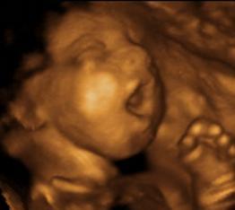
What does the 20-week scan show?
The anomaly scan checks your baby for structural abnormalities. The sonographer will check your baby’s head, face, spine and see whether all the bones align. All internal organs will be checked to see that they have developed properly. The heart is checked to ensure the four chambers are of equal size and the valves appear to be working with every heartbeat. Your baby’s kidneys and stomach will be checked to see if they’re functioning properly, and limbs, hands and feet will also be examined.
The position of the placenta will be noted; if it is lying low in your uterus you will need to have another scan later on. The sonographer will also check the umbilical cord and the volume of amniotic fluid surrounding your baby.
There will be various measurements made during the scan, mainly: Head circumference, Abdominal circumference, thigh bone length. These measurements indicate whether your baby is developing as expected and act as a double-check on your estimated due date.
As well as developmental checks, the sonographer is looking for specific conditions that may be treatable, or may jeopardise your baby’s survival. One has to note that certain anomalies like those involving spinal cord, brain, heart and kidneys have high chance of being picked up (80-90%), while some like bowel atresia do not manifest until late. Some may not be detectable before birth (hypospadias)
If the sonographer detects any problems, you will be referred to a fetal medicine specialist and should be seen within a few days. You will be examined again and it may be that your baby can be treated within the womb or that they will need treatment after their birth. In a few instances, you may be given the option to end your pregnancy. You will be given counseling and support when making this tremendously difficult decision.
The ability to check the baby’s development is dependent upon many factors. The bodyweight of the mother and the manner in which the baby lies in the womb are major issues. However, sometimes perfect views of a certain part of the baby’s body cannot be obtained. In this case you will be invited back for a repeat scan. This happens in about 10% of cases so please do not be alarmed if this happens to you.
One should understand that no prenatal test can detect all forms of birth defects and abnormalities.
Birth Defects: Prevention and Treatment
Congenital abnormalities can be divided into three groups:
- Lethal if the defects (such as anencephaly or hypoplastic left heart syndrome or bilateral renal agenesis) cause stillbirth or infant death; pregnancies are terminated after the prenatal diagnosis of fetal defects in more than 50% of cases.
- Severe if the defects (such as spina bifida, bladder exstrophy) without medical intervention cause handicap or jeopardize life. Parents are counseled regarding handicaps, and need for multiple hospital admissions for treatments/ procedures in children. The have an option to terminate the pregnancy.
- Mild if defects (such as unilateral hydronephrosis, cleft lip) require medical/surgical intervention but life expectancy is good.
The causes of congenital abnormalities
Genetic: which includes chromosomal aberrations (e.g. Down syndrome) Mainly two conditions may contribute to a higher total prevalence of congenital abnormalities with genetic origin: (i) women giving birth after 35 years of age and (ii) high rate of consanguineous marriages.
Environmental: which includes infectious diseases (e.g. rubella), maternal diseases (e.g. diabetes mellitus or diseases with high fever), teratogenic drugs, alcohol, smoking and environmental pollutants. The proportion of environmental origin may be about 15% of total congenital abnormalities.
Complex (multifactorial) origin: caused by gene-environmental interaction when the so-called polygenic liability (predisposition) is triggered by environmental ‘risk’ factors. Most common congenital abnormalities (such as isolated neural-tube defects, cleft lip, cardiovascular malformations, , congenital dislocation of the hip, hypospadias, etc) belong to this etiological group. The proportion of complex origin is estimated about 60% of total congenital abnormalities.
Prevention of congenital anomalies:
1. Primary prevention: avoiding the cause(s) of congenital abnormalities, e.g. rubella vaccination or periconceptional folic acid/multivitamin supplementation.
2. Secondary prevention: early detection followed by effective early treatment, e.g. neonatal orthopedic screening is very effective for the early detection and treatment of deformities such as congenital dislocation of the hip
3. Tertiary prevention: complete recovery of congenital abnormalities by early surgical intervention without residual defects or minimal after effects.
Consanguinity (relationship by blood)
Increases the prevalence of rare genetic congenital anomalies and nearly doubles the risk for neonatal and childhood death, intellectual disability and serious birth anomalies in first cousin unions.
Infections
Maternal infections such as syphilis and rubella are a significant cause of birth defects in low- and middle-income countries.
Maternal nutritional status
Iodine deficiency, folate insufficiency, overweight, or conditions like diabetes mellitus are linked to some congenital anomalies. For example folate insufficiency increases the risk of having a baby with neural tube defects.
Environmental factors
Maternal exposure to pesticides, medicinal and recreational drugs, alcohol, tobacco, certain chemicals, high doses of vitamin A during the early pregnancy, and high doses of radiation increase the risk of having a baby with congenital anomalies.
Prevention
Preventive public health measures administered through pre- and peri-conception and prenatal health care services decrease the frequency of certain congenital anomalies. Primary prevention of congenital anomalies involves:
- Improving the diet of women throughout their reproductive years, ensuring an adequate dietary intake of vitamins and minerals such as folic acid and iodine, and restricting harmful substances, particularly the abuse of alcohol. Controlling pre-conceptional and gestational diabetes through counselling, weight management, diet and the administration of insulin when needed.
- Avoiding exposure to hazardous environmental substances (e.g. heavy metals, pesticides, some medicinal drugs) during pregnancy.
- Improving vaccination coverage, especially with rubella virus, for children and women. This can be prevented through childhood vaccination. The rubella vaccine can also be given at least 1 month prior to pregnancy to women who are not already immune.
- Increasing and strengthening education to health staff and others interested in promoting birth defects prevention.